
Introduction:
Microscopy in the laboratory is the field of imaging biological specimens (cells, tissues, and biomolecules) using all forms of instrumentation/techniques to obtain a detailed picture (both at the mesoscale and at the nanoscale); therefore, includes all forms of microscopy. From basic brightfield light microscopes to high-end confocal and super-resolution systems, laboratory imaging microscopy is central to cell biology, pathology, and materials science. LabXcite, laboratory equipment supplier in Bangalore, Karnataka, India, supports laboratory imaging microscopy instrument selection and installation for Karnataka’s life sciences research community.
What Is Laboratory Imaging Microscopy?
Laboratory imaging microscopy is the practice of using optical or electron-based systems to produce magnified, high-resolution images of samples at the cellular and subcellular level. The range of laboratory imaging microscopy is enormous — from a simple brightfield microscope used in a teaching lab to a STED super-resolution system that can image protein complexes below 50 nm. Research institutions in Bangalore like NCBS, IISc, and IBAB use advanced laboratory imaging microscopy for cell biology, structural biology, and neuroscience research that feeds into India’s growing biotech sector.
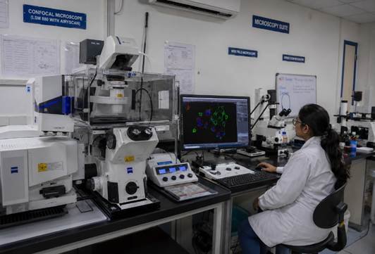
Types of Microscopy Techniques in Research Labs
Laboratory imaging microscopy spans eight main technique categories. Knowing which one fits your biological question saves significant time and budget:
| Microscopy Type | Resolution Limit | Sample Type | Best Application | Relative Cost |
| Brightfield | ~500 nm practical | Fixed, stained | Routine histology, stained cell monolayers | Low |
| Phase Contrast | ~200–500 nm | Live or unstained | Live cell morphology without staining | Low–Medium |
| Widefield Fluorescence | ~200 nm | Fixed or live, labelled | Protein localisation, organelle imaging | Medium |
| Confocal (Point-Scan) | ~200 nm lateral | Fixed or live (short) | 3D optical sectioning of thick specimens | High |
| Spinning Disk Confocal | ~200 nm | Live cells (long time-lapse) | Extended time-lapse, low phototoxicity | High |
| TIRF | ~100 nm axial | Live cells at coverslip surface | Membrane receptor dynamics | High |
| SEM / TEM (Electron) | 0.1–5 nm | Fixed, dehydrated | Ultrastructure, nanoparticle imaging | Very High |
| Super-resolution (STED/STORM) | 20–80 nm | Fixed (STORM) or live (STED) | Protein complexes below diffraction limit | Very High |
Laboratory imaging microscopy selection always starts with your resolution requirement and sample type — not with the most impressive instrument specification available.
Fluorescence vs Brightfield Microscopy
Brightfield laboratory imaging microscopy is the right choice when your sample has inherent contrast from staining — H&E for tissue sections, crystal violet for bacteria, Gram stain for clinical microbiology. Fluorescence laboratory imaging microscopy is needed when you want to localise a specific protein, track a molecule over time in a living cell, or image multiple targets simultaneously with different colours. The trade-off is time — fluorescence labelling adds sample preparation steps — but the specificity you gain is often irreplaceable.
| Feature | Brightfield Microscopy | Widefield Fluorescence Microscopy |
| Contrast source | Stain or pigment absorption | Fluorescent label emission |
| Sample prep time | 30 min–1 hr staining | 1–24 hrs labelling with dye or antibody |
| Specificity | Morphological only | Target-specific — protein, organelle, nucleic acid |
| Live cell compatible | Yes (unstained) or stained fixed | Yes, short-term with live dyes or fluorescent proteins |
| Equipment cost | Low | Medium to High |
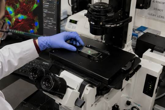
Live Cell vs Fixed Cell Imaging
Choosing between live and fixed laboratory imaging microscopy depends on your biological question:
Live cell laboratory imaging microscopy:
- Captures real biological dynamics — division, migration, protein trafficking, cell death.
- Requires an environmental control unit (37°C, 5% CO + humidification) when working with cells from mammals.
- The risk of phototoxicity limits how long and how bright the specimen can be illuminated.
- Shows biology as it actually happens — not a preserved, fixed snapshot.
Fixed cell laboratory imaging microscopy:
- No environmental control needed during imaging — simpler setup.
- Multi-step antibody labelling possible — reveals multiple targets simultaneously.
- Higher resolution possible — longer imaging times with no phototoxicity concern.
- Cannot capture dynamics — you see one frozen moment in time.
How to Choose the Right Microscopy System for Your Lab
- Start with your biological question. Do you need to localize a specific protein? Image a whole tissue section? Track cell movement over 24 hours? The answer to this question eliminates most options before you even look at a catalogue for laboratory imaging microscopy systems.
- Determine your resolution requirement. Most cell biology questions are answered at widefield fluorescence resolution (~200 nm). You only need confocal or super-resolution laboratory imaging microscopy if your structures are small enough that out-of-focus background genuinely compromises your data.
- Plan for live vs fixed from the start. Live cell laboratory imaging microscopy requires a stage incubator, a CO2-compatible enclosure, and a spinning disk confocal if extended time-lapse is planned. Budget and specify this infrastructure before ordering the microscope body.
Frequently Asked Questions
Q: What are the main types of microscopy used in research labs?
A: The main types include brightfield (stained cells and tissue), widefield fluorescence (labelled proteins and organelles), confocal (3D optical sectioning), spinning disk confocal (live cell time-lapse), electron microscopy SEM/TEM (nanometre resolution ultrastructure), and super-resolution microscopy STED/STORM for imaging below the 200 nm diffraction limit.
Q: What is the difference between light and electron microscopy?
A: Light microscopy uses visible light to obtain a resolution of approximately 200 nanometers, which is adequate to display the morphology of most cellular structures (e.g., many of the cells in a multicellular organism). By using a focused electron beam to obtain a resolution of 0.1 to 1.0 nanometers, it is possible to visualize many of the structures within the cell, including ribosomes and membrane sub-complexes. Live image acquisition can be accomplished using light microscopy; electron microscopy requires the specimen to be fixed and dehydrated prior to imaging.
Q: What is the best microscope for imaging live cells?
A: Live cell imaging is often done using widefield fluorescence microscopy because of its fast acquisition speed. When multiple fields are being imaged over an extended period of time, spinning disk confocal microscopy is superior because of its ability to reduce phototoxic effects. For larger 3D specimens such as organoids, light sheet microscopy is quickly becoming an optimal choice.
Q: What is the difference between confocal and widefield fluorescence microscopy?
A: When imaging using widefield, light illuminates the entire area at one time; although it can obtain images quickly, it does create out of focus backgrounds when viewing through thick samples. In contrast, confocal utilizes a pinhole to eliminate out-of-focus light, which produces clearer images from thick samples and allows for reconstruction in 3D space. In addition, confocal systems are far more expensive and slower than widefield systems for each frame acquired.
Conclusion
Laboratory imaging microscopy is one of the most powerful ways to understand biology — seeing structures, dynamics, and interactions that cannot be inferred from bulk measurements alone. Choosing the right laboratory imaging microscopy platform requires matching technique to biological question, budget to genuine need, and supplier to service capability. LabXcite, laboratory equipment supplier in Bangalore, India, can help you navigate the laboratory imaging microscopy options for your research context. Visit products to explore imaging and microscopy equipment.