
Introduction:
Cell culture techniques are methods that enable the growth, maintenance, and manipulation of animal, plant or microbial cells outside of their natural biological environment. They occur within controlled laboratory conditions. When researchers employ core cell culture techniques, such as sterile handling of the materials, managing media correctly, subculturing, assessing the viability and contamination prevention, labs produce reproducible results that can be used in biomedical research, drug testing and vaccine development. LabXcite is a laboratory equipment supplier located in Bangalore, Karnataka, India. They offer the same cell culture technique equipment to Biocon, Mynvax, Strand Life Sciences and research labs throughout Karnataka.
What Is Cell Culture in Biology?
The purpose of the media in which cells are grown is to simulate physiological conditions outside of a living organism. These cell culture techniques can use established immortalised cell lines — like HeLa, HEK293, or CHO cells — that grow indefinitely in culture, primary cells freshly isolated from tissue with limited lifespan, or stem cells requiring specialised growth factors and substrates. Cell culture techniques underpin most modern biomedical research, biopharma manufacturing (antibodies, vaccines, recombinant proteins), and drug compound screening. India’s biopharma sector, anchored by Bangalore companies like Biocon, is investing heavily in expanding cell culture techniques capacity for biologics and cell therapy manufacturing.
Essential Cell Culture Equipment
These five instruments are non-negotiable for any mammalian cell culture techniques lab:
| Equipment | Function in Cell Culture Techniques | Key Specification |
| Class II Type A2 Biosafety Cabinet | Provides sterile, HEPA-filtered working environment for all cell handling — protects cells and operator simultaneously | HEPA ≥99.97% efficiency; ≥0.45 m/s inward airflow at sash; DEHS aerosol challenge certification |
| CO2 Incubator | Maintains 37°C, 5% CO2, and 95% humidity — the three parameters mammalian cells need for physiological growth | ±0.1°C uniformity; IR CO2 sensor preferred; copper or copper-alloy interior for antimicrobial protection |
| Inverted Phase Contrast Microscope | Daily cell health monitoring — confluency, morphology, contamination check without disturbing the culture | 10× and 20× phase contrast objectives; camera port for documentation |
| Refrigerated Benchtop Centrifuge | Cell pelleting, washing, and passaging at 300–400 × g without damaging cell membranes | Swing-bucket rotor for 15/50 mL tubes; temperature control 4–37°C |
| Cell Counter (Auto or Manual) | Counting cells and assessing viability before seeding at defined density | Trypan blue exclusion for routine use; fluorescence dual-stain for low-viability samples |
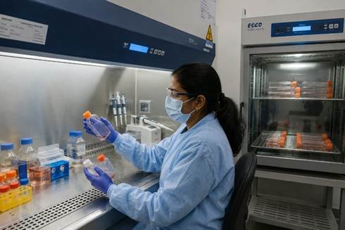
Basic Cell Culture Techniques — Step-by-Step Passaging Protocol
Passaging or subculturing is the most frequently performed of all cell culture techniques in any mammalian cell biology lab:
- Prepare your Class II biosafety cabinet: UV-sterilise for 15 minutes, wipe all interior surfaces with 70% ethanol, switch on the blower and allow air to stabilise for 10 minutes before starting cell culture techniques work.
- Warm complete culture media and PBS to 37°C. Cold media will create osmotic pressure on the cell and delay recovery from the cell culture techniques that were performed on it.
- Check the confluence of the cells you are going to use under a microscope. The confluence should be between 70 – 80% and cells should look normal and not contaminated (cloudy).
- After checking the confluence of the cells, aspirate off the old media and wash the cells once with PBS. This removes any residual serum proteins in the media that would affect the function of trypsin in the following step of cell culture.
- Add pre-warmed 0.25% trypsin-EDTA. Incubate 2–5 minutes at 37°C, watching under the microscope until cells round up and detach. Never over-trypsinise — this is one of the most common errors in mammalian cell culture techniques.
- Neutralise trypsin by adding 3× volume of complete media. When making a cell suspension, pipette gently to make a single-cell suspension — do not pipette vigorously as this will damage the cells and decrease viability.
- Count the number of cells and calculate the seeding density to determine the correct number of cells to seed into the new flasks. Seeding a cell density outside the determined range will lead to inconsistencies between passages.
- Return flasks to the CO2 incubator and record passage number, seeding density, date, and operator in your cell culture techniques logbook.
Common Cell Culture Problems & Troubleshooting
Contamination is the single biggest source of failed cell culture techniques experiments. Here is how to identify and respond to the four most common problems:
| Problem | Visual Signs | Probable Cause | Solution for Cell Culture Techniques Lab |
| Bacterial contamination | Media turns cloudy or turbid; pH drops rapidly (yellow in phenol red media); visible particles | Break in aseptic technique; contaminated reagent; non-sterile equipment | Discard all affected flasks. Decontaminate BSC. Identify and fix the contamination source before restarting cell culture techniques. |
| Fungal contamination | Visible fluffy or strand-like growth in media; gradual pH drop | Environmental spores entering BSC; contaminated media | Discard all cultures. Deep-clean BSC and incubator interior. Check media sterility and incubator water pan. |
| Mycoplasma contamination | No visible signs — cells grow slowly, behave unexpectedly in assays, have unusual gene expression | Contaminated cell line from external source; contaminated serum | Test all cell lines quarterly by PCR or ELISA. Discard confirmed positives. Quarantine all new lines before use in cell culture techniques. |
| Poor growth or senescence | Slow growth, failure to reach confluency, premature detachment, abnormal morphology | High passage number; poor media quality; CO2 or temperature drift | Check incubator calibration. Verify media freshness. Return to low-passage frozen stock. These are common signs your cell culture techniques setup needs recalibration. |
Mycoplasma is the most dangerous contamination in cell culture techniques labs because it is completely invisible to the naked eye — detectable only by PCR or ELISA — and silently alters cell metabolism and gene expression for months before anyone suspects a problem.
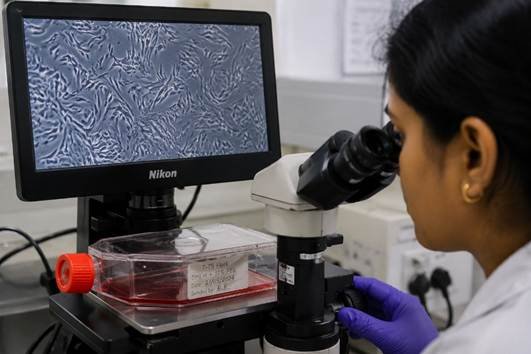
How to Prevent Contamination in Cell Culture
- Always work inside a properly certified Class II BSC with validated airflow. Cell culture techniques performed on an open bench — even ‘just briefly’ — are almost certain to fail.
- Test every new cell line for mycoplasma before using it in any cell culture techniques experiment. This single rule prevents the most costly and hard-to-detect contamination in any cell biology lab.
- Use only sterile-filtered media and reagents from quality-tested suppliers. If you prepare your own media, filter through a 0.2 µm membrane immediately before use in cell culture techniques.
- Dedicate one media bottle to each cell line. Never pour from a shared reservoir into multiple cell culture techniques flasks — shared bottles are a contamination path between cell lines.
- Replace the CO2 incubator water pan weekly with fresh sterile distilled water. The water pan is a major source of fungal and bacterial contamination in cell culture techniques facilities.
- UV-sterilise the BSC before every session and wipe with 70% ethanol after every session. A clean BSC is the single most effective contamination prevention measure in all cell culture techniques.
- Change gloves between handling different cell lines. When transferring media, use a new pipette tip for each transfer. Using a shared consumable between lines will be a source of contamination for your cell culture.
- Use pre-warmed media at 37°C. Adding cold media to the cells will cause thermal shock and reduce viability and increase recovery time. This is a common but overlooked principle of good cell culture practice.
Frequently Asked Questions
Q: What is cell culture in biology?
A: Growing Animal, Plant, or Microorganism Cells Outside Their Natural Habitat in an Artificial Medium Under Controlled Temperature, Humidity, & CO2 Amount; e.g. 37oC (Mammalian Cells), 5% CO2 and 95% RH) Infers Research; Drug Screening and Development; Production of Biomolecules; Creation of Models (Disease, etc… ) Cell Culture is Foundational to: Producing Vaccines; Researching Tumors; Pharmaceutical Manufacturing.
Q: How do you perform cell culture step by step?
A: Basic Passaging Sequence: Prepare Sterile Class II BSC; Warm Media and PBS to 37oC; Examine Cells with Inverted Microscope for Degree of Confluency & Health – From Above Perspective; Remove Old Media by Aspirating; Wash Cells with Phosphate Buffered Saline (PBS); Add Trypsin; Incubate 2-5 Minutes (37 o C); Neutralize with Complete Media; Count Cells; Seed Cells at Desired Density; Return to CO2 Incubator. Record passage number, seeding density, and date in your cell culture techniques logbook.
Q: What are the most common cell culture contamination problems?
A: The most common are bacterial (visible turbidity and pH drop from aseptic technique break), fungal (visible strand-like growth from environmental spores), and mycoplasma (completely invisible, detected only by PCR or ELISA, silently alters cell behaviour for months). Mycoplasma is the most dangerous contamination in cell culture techniques because it is undetectable without specific testing.
Q: How do you prevent contamination in mammalian cell culture?
A: Key prevention: always work in a certified Class II BSC, test every new cell line for mycoplasma before use, use sterile quality-tested media, dedicate media bottles per cell line, replace the incubator water pan weekly, UV-sterilise the BSC before every session, change gloves between cell lines, and always use pre-warmed media. These cell culture techniques contamination prevention rules eliminate the vast majority of contamination events.
Conclusion
Reliable cell culture techniques take practice, discipline, and the right equipment — but once mastered, they unlock the most powerful experimental systems in modern biology and biopharma. The cell culture techniques contamination problems that most commonly derail experiments — bacterial, fungal, and mycoplasma — are all preventable with consistent aseptic practice, regular testing, and properly maintained equipment. LabXcite, laboratory equipment supplier in Bangalore, India, supplies cell culture techniques equipment and consumables — CO2 incubators, biosafety cabinets, culture media, and pipette tips. Visit products for the full cell culture range.