
Introduction:
Live cell imaging is the use of fluorescence microscopy to visualize changes and movements of living cells as they take place, including the processes of cell division and motility. Good live cell imaging practice entails a careful management of temperature, illumination, and time in order to ensure optimal cell cultivation with regard to subsequent experimental processes.
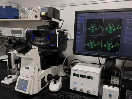
What Is Live Cell Imaging?
Live cell imaging enables observing processes as they occur, instead of relying on just one fixed point in time. Questions about speed, movement, and timing can only be answered through cell imaging, since fixed images freeze a single moment and cannot show change over time. Bangalore and Karnataka research institutes increasingly rely on live cell imaging to study cell behaviour in real time.
Essential Equipment for Live Cell Imaging
A spinning disk confocal system is also a strong option for live cell imaging when you need long time-lapse sessions with less light damage to cells.
| Equipment | Why It Matters |
| Inverted fluorescence microscope | Lets the lens view cells from below the dish |
| Environmental chamber | Keeps 37°C, CO2, and humidity stable |
| Sensitive camera | Captures clear images at low light levels |
| Automated stage | Allows imaging of many positions at once |
Fluorescent Labels Used in Live Cell Imaging
Choosing the right label is one of the first decisions in planning good live cell imaging, since some dyes are more toxic to cells than others over long sessions.
| Label Type | Best Use |
| Fluorescent proteins (GFP, mCherry) | Genetically tagging target proteins |
| Live cell dyes | Staining organelles without transfection |
| Fluorescent antibody fragments | Labelling surface receptors |
| Quantum dots | Bright, long-lasting signal |
Controlling Phototoxicity
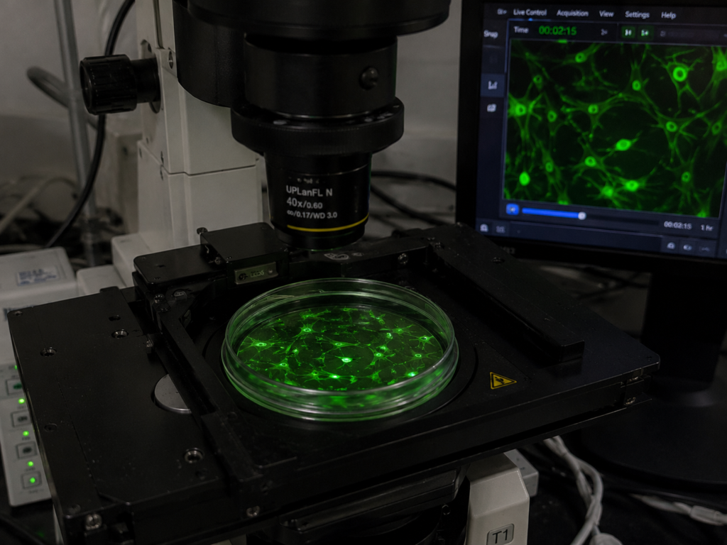
Too much light is the biggest risk in live cell imaging, since excess light energy can quickly damage or kill the very cells you are trying to study.
- Use the lowest light power that still gives a clear image.
- Space out imaging intervals instead of shooting continuously.
- Choose red-shifted fluorophores where possible.
- Increase the sensitivity of the camera instead of light intensity.
- Introduce antioxidants into the culture.
- Use spinning disk confocal during long procedures.
Setting Up Time-Lapse Acquisition
- Minimize the exposure time.
- Choose a time interval that will allow capturing the process without excessive exposure to the cells.
- Set a reasonable z-stack range for having a good Vo2.
- Establish total duration of the experiment beforehand.
- Select a reliable autofocus method.
FREQUENTLY ASKED QUESTIONS
Q: What instruments do I need to use for live cell imaging?
A: You need an inverted fluorescence microscope, an on-stage environmental chamber to hold temperature and CO2 steady, a sensitive camera for low-light detail, and glass-bottomed dishes suited for live cell imaging. A spinning disk confocal is a helpful upgrade for long time-lapse sessions.
Q: What is the way to minimize the phototoxicity while doing live imaging?
A: One can achieve optimum live cell imaging results by providing the lowest light that still allows for identifiable images, increasing the time intervals in between imaging sessions and the use of red fluorescent tags. All of these techniques reduce injury caused by light to the cells under observation.
Q: What is the duration of cell viability in live cell imaging?
A: If temperatures and CO2 concentrations are kept at appropriate levels and exposure to light minimized, healthy cells can remain alive during live cell imaging experiments for 12-72 hours. Poorly controlled light exposure, however, can cause cell stress or death within just a couple of hours.
Q: What type of microscope is appropriate for live cell imaging?
A: A wide-field inverted fluorescence microscope with an environmental chamber is suitable for live cell imaging. When time-lapse imaging is to be conducted for a long duration of time, spinning disk confocal is preferred to minimize light exposure while creating clear and informative images.
Conclusion
Successful live-cell imaging requires using the correct equipment, ensuring effective light control, and careful design of experiments that help collect accurate information about cell functioning by preserving cell health. Are you planning to set up a live-cell imaging system? LabXcite is the most reputed supplier of scientific laboratory equipment in Bangalore, India. You can browse through their wide range of products.