
Introduction:
The microtome sectioning technique refers to the use of techniques to cut tissue samples into thinner cuts for viewing through a microscope. These techniques are important in pathology laboratories, research units, and examination areas. Efficient microtome sectioning techniques enable clear and uniform sections that allow clinicians and researchers to accurately examine the tissues, hence allowing them to make correct diagnoses and reliable research outputs.
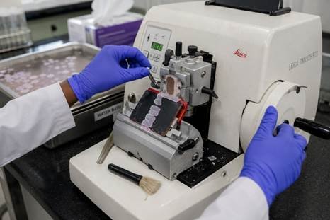
What Is a Microtome?
A microtome is a special kind of equipment that advances the tissue piece by small distances each time, which is usually in micrometers and then cuts it. Different microtome slicing methods are applicable for different jobs, ranging from the most ordinary hospital slides to the more complicated electron microscope samples. Pathology units and diagnostic laboratories in Bangalore use various techniques simultaneously as per the particular type of material and speed of the result needed to be obtained.
Types of Microtomes
| Microtome Type | Thickness Range | Main Use |
| Rotary microtome | 3-5 µm | Paraffin sections, routine slides |
| Cryostat | 5-10 µm | Frozen sections, fast diagnosis |
| Vibratome | 40-100 µm | Fresh unfixed tissue, brain studies |
| Sledge microtome | Varies | Large samples like bone |
| Ultramicrotome | 60-100 nm | Electron microscope prep |
Choosing the Right Section Thickness
Different services need different thicknesses. Routine tissue slides usually need 3-5 µm sections. Special stains may need slightly thicker slices, around 5-6 µm. Choosing the correct thickness is one of the most important microtome sectioning techniques a technician learns early in training, since sections that are too thick can hide important cell details from the pathologist.
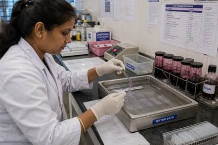
Basic Steps for Cutting Paraffin Sections
Following these steps in order is one of the most reliable microtome sectioning techniques for producing clean, ribbon-like paraffin sections every time.
- Chill the tissue block on ice.
- Remove extra wax by trimming the block.
- Place the blade and fix it.
- Adjust the slicing thickness properly.
- Cut a clean ribbon of sections.
- Float the ribbon on a warm water bath.
- Lift sections onto a glass slide.
- Drain and dry the slide in an oven.
Fixing Common Section Problems
Most cutting problems trace back to temperature, blade sharpness, or cutting speed, and fixing these quickly is a core part of good microtome sectioning techniques.
| Problem | Likely Cause | Fix |
| Chattering lines | Cutting too fast | Slow down cutting speed |
| Folding | Tissue too warm | Chill block before cutting |
| Fragmenting | Wax too cold | Adjust room or block temperature |
| Thick sections | Wrong calibration | Check advance setting |
| Blade lines | Damaged blade | Change to a fresh blade |
FREQUENTLY ASKED QUESTIONS
Q: What are the main types of microtomes used in histology?
A: The main types are the rotary microtome for routine paraffin slides, the cryostat for fast frozen sections, the vibratome for fresh unfixed tissue, the sledge microtome for large samples, and the ultramicrotome for electron microscope work.
Q: What section thickness should I use for routine histology?
A: Most routine histology slides are cut at 3-5 micrometres. Thicker sections can cause cells to overlap and make details harder to see clearly under the microscope.
Q: How do I prevent sections from folding or chattering?
A: Chill the tissue block on ice for a few minutes before cutting to stop folding, and slow down your cutting speed to reduce chattering. A fresh, sharp blade also fixes most cutting problems quickly.
Q: What is the difference between a cryostat and a rotary microtome?
A: A rotary microtome cuts tissue that has been processed and set in wax, giving very detailed slides but taking longer to prepare. A cryostat cuts fresh frozen tissue in minutes, which is useful during surgery when a fast result is needed.
Conclusion
Learning the right methods of microtome slicing requires practice but is worth it since it allows making high-quality slides every time. Whether you work with paraffin blocks or frozen tissue, the right technique and equipment make all the difference to diagnosis quality and research accuracy. Need a microtome or histology accessories for your lab? LabXcite, laboratory equipment supplier in Bangalore, India, offers trusted histology equipment with local support – check our products today.